Atlas of Regional, Functional and Radiological Anatomy
VOXEL-MAN 3D Navigator Brain and Skull is a very useful introduction to the morphology, functional anatomy, and blood supply of the brain. The integrated approach and 3D interactive format are user-friendly and highly suitable for the target audience. — Jacqueline Graham and Paul D. Griffiths, Neuroradiology (more reviews)
VOXEL-MAN 3D Navigator: Brain and Skull is an new kind of anatomical and radiological atlas. It is novel in at least two respects:
- Unlike books or traditional multimedia programs, it allows interactive exploration of a detailed three-dimensional anatomy model. Each structure is labeled and described, and can thus be interrogated directly on the screen. The advantages of dealing with real anatomy are thus combined with the advantages of learning from a book (associated knowledge).
- Unlike traditional sources of knowledge, VOXEL-MAN 3D Navigator presents the radiological manifestation of normal anatomy in the context of three-dimensional anatomy. It thus decisively improves the understanding of both X-ray and cross-sectional radiological images.
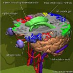 VOXEL-MAN 3D Navigator: Brain and Skull is based on various virtual body models, created from cross-sectional images from computed tomography (CT) and magnetic resonance imaging (MRI). In addition, images from the Visible Human were used. It contains more than 250 three-dimensional anatomical objects, including functional areas, vessels, and blood supply areas.
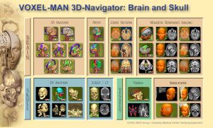 The anatomy is presented in 36 interactive scenes that are accessed from a visual table of contents and can be explored and dissected in various ways. The interactive scenes cover a variety of topics such as subdivision of the cerebral cortex, transverse MRI cross-sections, exploded view of the skull, and blood vessels of the head. 18 of the scenes are also available in stereoscopic (red/green) format.
 Using the mouse, the scene may be rotated, and objects may be added or removed. Furthermore, anatomical objects may be marked, searched for, painted, annotated, or described. Anatomical objects may be selected in the image or from an object list. A textual instruction illustrates the possibilities available for the chosen scene.
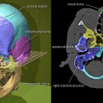
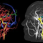 The radiological manifestation of the objects can be viewed in the context of 3D anatomy. This includes simulated X-ray images, as well as maximum intensity projections and cross-sectional images from CT and MRI.
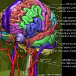 VOXEL-MAN 3D Navigator: Brain and Skull is a multilingual system: The anatomical nomenclature is available in Latin, English, French, German, and Japanese (French and Japanese only for the brain without vascular system). For English, German and Japanese Windows systems, the user interface automatically adapts to the respective language. A manual is available in English and German.
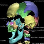 The program provides unique reference material not only for medical students, but also for professionals in all medical disciplines involving anatomy and radiology of the head. Even laypersons can get fascinating insights.
Editions
![]() VOXEL-MAN 3D Navigator: Brain and Skull was first published in 1998 by Springer-Verlag (as VOXEL-MAN Junior: Brain and Skull), with updated versions published in 2001 and 2009, respectively. Since 2018, it is available for free download under a Creative Commons license. A Chinese version was published in 2002 by Zhejiang University Press.
VOXEL-MAN 3D Navigator: Brain and Skull was first published in 1998 by Springer-Verlag (as VOXEL-MAN Junior: Brain and Skull), with updated versions published in 2001 and 2009, respectively. Since 2018, it is available for free download under a Creative Commons license. A Chinese version was published in 2002 by Zhejiang University Press.
References
- Karl Heinz Höhne, Andreas Petersik, Bernhard Pflesser, Andreas Pommert, Kay Priesmeyer, Martin Riemer, Thomas Schiemann, Rainer Schubert, Ulf Tiede, Markus Urban, Hans Frederking, Mike Lowndes, John Morris: VOXEL-MAN 3D Navigator: Brain and Skull. Regional, Functional and Radiological Anatomy. Version 2.0. Springer-Verlag Electronic Media, Heidelberg, 2009. DVD, ISBN 978-3-642-01211-2.
Back to VOXEL-MAN 3D Navigators