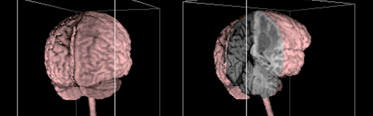
At the 73rd Scientific Assembly and Annual Meeting of the Radiological Society of North America (RSNA) in Chicago, we presented the first ever 3D reconstruction of the complete brain of a living person as a poster and a video.
The underlying 3D model is based on two image datasets from magnetic resonance imaging (MRI). Each dataset consists of 128 sagittal cross-sectional images with a resolution of 256 x 256 pixels. They were acquired with a Siemens Magnetom scanner, using Fast Low Angle Shot Imaging (FLASH) and Fast Imaging with Steady-state Precession (FISP) MRI sequences, respectively.
 The image processing environment was implemented on a DEC VAX-11/780 minicomputer under VMS with two connected image workstations (COMTAL Vision One/20, VTE Picturecom). Detection of the contours of the brain using a 3D Marr-Hildreth operator (3D Mexican hat) took about 230 minutes of computing time, followed by about 45 minutes of interactive correction of some erroneous bridges between brain and soft tissue. The 3D images of the brain were produced with the VOXEL-MAN-8 program. Calculating a single 3D image (right) from the 3D brain model again required several minutes of computing time.
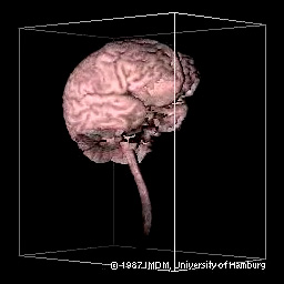
Here are two short sequences from the original digital video. A somewhat longer section can be found in the video Visualizing the Human Body.
In the video, the flattening of the gyri in the right hemisphere caused by a tumor is clearly visible. Various sections illustrate the location of the metastasis (blue) and a surrounding edema. The original gray values from FLASH and FISP images, as well as the first principal component of the Karhunen-Loeve transform (KL-TR), are mapped to the cut planes, emphasizing different aspects of the lesion.
This work was awarded a DAGM Honorable Mention by the German Association for Pattern Recognition. It provided the foundation for later, considerably refined 3D brain models and atlases.
References
- Michael Bomans, Karl Heinz Höhne, Ulf Tiede, Martin Riemer: 3-D segmentation of MR images of the head for 3-D display. IEEE Transactions on Medical Imaging 9 (2), 1990, 177-183.
- Michael Bomans, Martin Riemer, Ulf Tiede, Karl Heinz Höhne: 3-D Segmentation von Kernspin-Tomogrammen. In Erwin Paulus (ed.): Mustererkennung 1987, Proc. 9. DAGM-Symposium, Informatik-Fachberichte 149, Springer-Verlag, Berlin, 1987, 231-235.
- Karl Heinz Höhne, Ulf Tiede, Martin Riemer, Michael Bomans, Martin Heller, Gerd Witte: Static and dynamic three-dimensional display of tissue structures from volume scans. Radiology 165 (P), 1987, 420.
Back to VOXEL-MAN Gallery