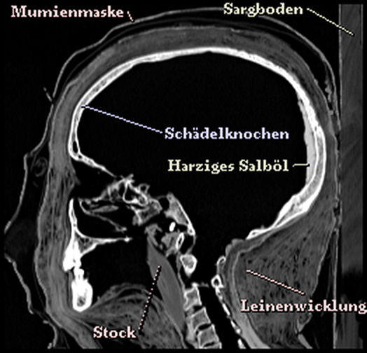
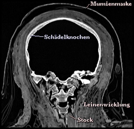
 A stack of transverse cross-sectional images from computed tomography (CT, left) forms the basic material for the Virtual Mummy. Various structures such as the light bone and the somewhat darker linen wrapping surrounding it are clearly visible. The sagittal (center) and coronal (right) cross-sectional images are reconstructed from this.

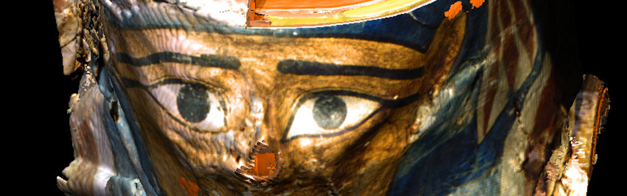
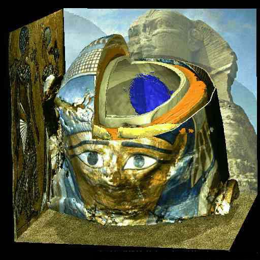
An image sequence of reconstructed perspective views simulates the unwrapping of the mummy. Four partially colored light sources serve as illumination. The light from below creates a somewhat ghosty impression.
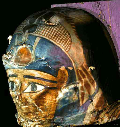
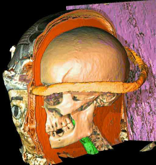
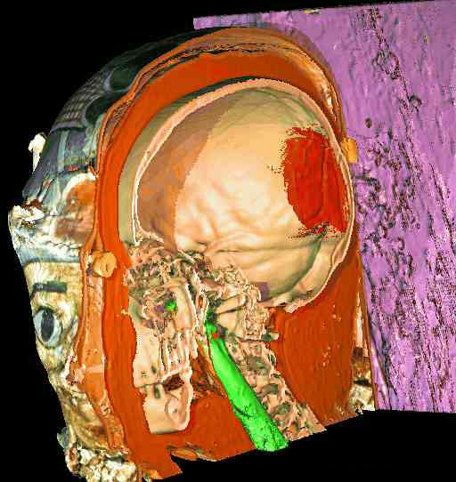
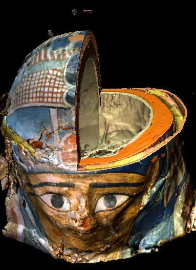
The head is covered by a painted mask, made of linen cardboard. Underneath lies the crown of justification (yellow). The skull is wrapped in several layers of linen (orange). At the back of the skull, traces of hardened ointment (red) can be found. A stick (green) serves as stabilization for the head. The mummy still rests in the original lower part of the 2300 year-old coffin (violet).
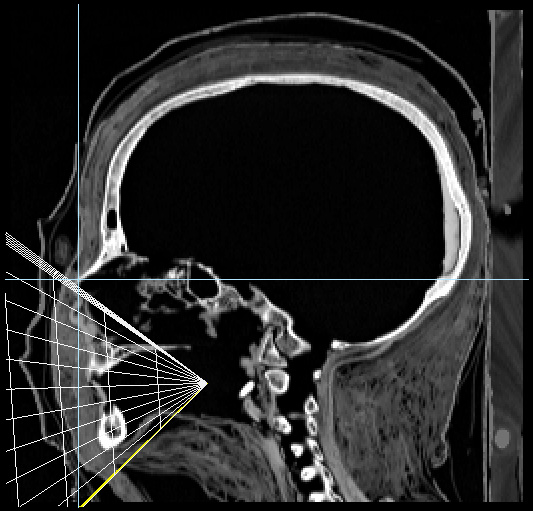
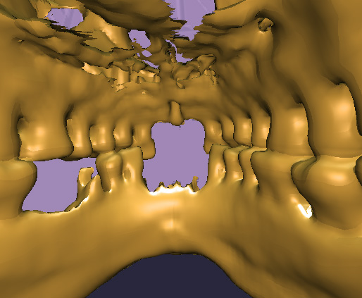
For the presentation of the Virtual Mummy, there are no limits to the imagination (left). Computer graphics also makes it possible to arbitrarily choose the position of the observer. For example, the virtual camera can be placed in the oral cavity (center), so that an inside view of the teeth is created (right).

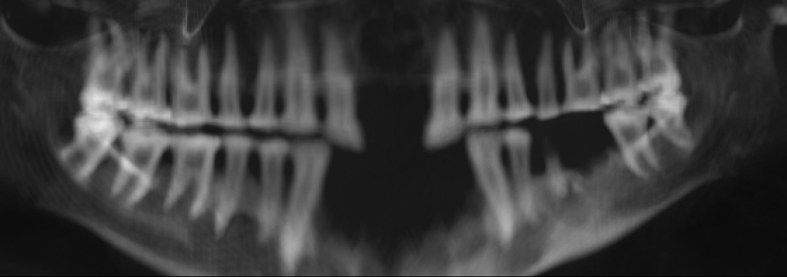
Panoramic Radiograph
The program can also calculate artificial X-ray images, for example a panoramic radiograph of the teeth of the mummy, similar to those commonly used by a dentist. It shows some interesting findings.
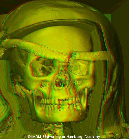
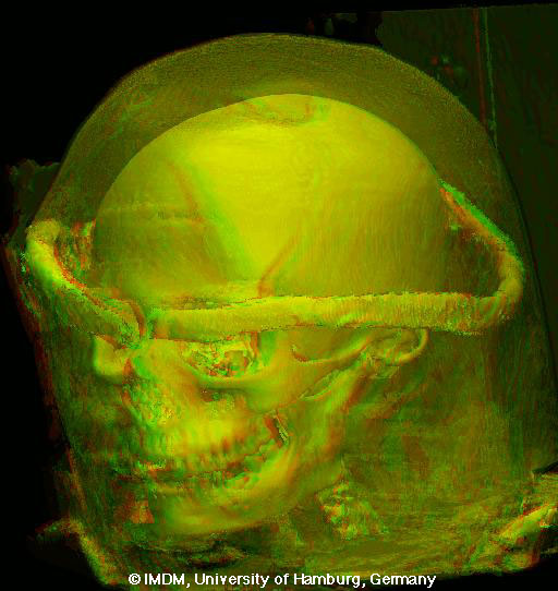
Red/Green Stereo
Depth perception can be enhanced by a stereogram of the image. These images must be viewed with red/green glasses. Colors are lost, though, when using this method.

See also body and winged scarab of other Egyptian mummies
Back to the Virtual Mummy